Treatment methods

Rehabilitation and physical therapy is a must for children with movement disorders. Ongoing medical monitoring and family support can significantly help children with this birth defect achieve the best quality of life possible.
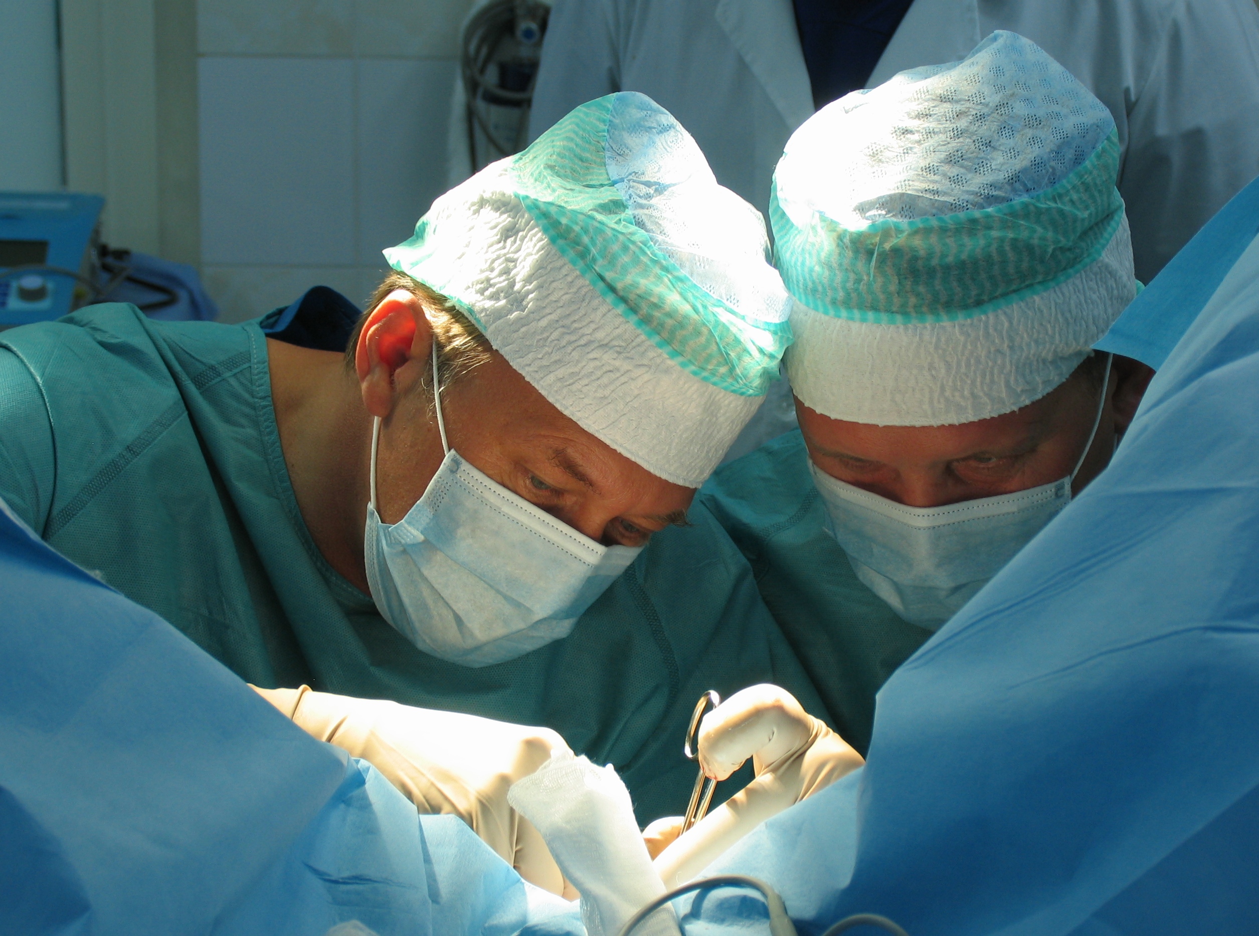
Children who have mild forms of this condition need only observation and care of the abnormal area. In a more serious manifestation of the disease, specialists resort to surgical intervention. Thus, they correct the defect and restore the natural anatomy.
The causes that lead to the development of spina bifida are not always known, but may include genetic factors and environment. For example, folic acid deficiency in early pregnancy can increase the risk of developing this birth defect. It is also important to consider family history of disease, as having cases of spina bifida in the family may increase the risk in offspring.
The symptoms of spina bifida can vary greatly depending on the severity of the abnormality. Mild forms of spina bifida may be asymptomatic or cause only minor symptoms.
Bulging of the skin or membrane over a defect on the baby’s back: This may be seen immediately after birth. In more serious cases where the spinal cord is also involved, paralysis of the lower limbs or other motor impairments may occur. Spina bifida can affect urinary control and cause other disorders in this area.
Diagnosis at MDI Clinic
Diagnostic measures usually begin with observation and visual inspection of the newborn. If there is a visible defect on the back or a bulge of skin or membrane over the back area, the doctor may immediately assume spina bifida.
Ultrasound can help detect signs of spina bifida in the fetus during pregnancy. However, this study may not always be sufficiently accurate and it is important to additionally perform other examinations after birth.
This method is more accurate for the diagnosis of spina bifida in newborns. MRI allows visualization of the structures of the spine and spinal cord, as well as evaluation of the degree of cerebral membrane presentation through the defect.
Fluoroscopy can sometimes be used to confirm the diagnosis, but it is a less preferred method because of the radiation of x-rays and visualization limitations.
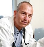
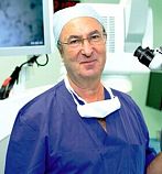
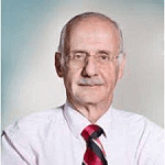
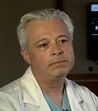
The best doctors in Israel
All doctorsPrice
How we are working
-
StepSubmitting an application

Simply leave a request or contact us at the numbers in the contact tab.
-
StepTalking to a counselor

You will be contacted by our consultant shortly after submitting your application. After the interview and review of the medical history, he will proceed to prepare a treatment program.
-
StepProgram preparation

Our specialists will draw up a personalized program, including a diagnosis and treatment schedule, the names and positions of the doctors, and the cost of treatment.
-
StepTravel arrangements

The coordinator will plan and organize the trip in every detail – from advice on preparing documents, to purchasing tickets, booking accommodation and even organizing excursions.
-
StepTreatment

Our staff will provide patient support throughout the diagnosis, treatment and rehabilitation period.

Форма обратной связи
"*" indicates required fields

